* Dipartimento di Informatica, Università di Pisa
** Dipartimento di Scienze Storiche del Mondo Antico - Egittologia, Università di Pisa
*** CNR (National Research Council) ITABC (Institute of Technologies Applied to Cultural Heritage)
**** CINECA-VISIT (Centro Interuniversitario di Supercalcolo - Laboratorio di Visualizzazione scientifica) , Casalecchio sul Reno Bologna
***** Dipartimento di Scienze Archeologiche - Antropologia, Università
di Pisa
The problem of rebuilding a face from human remains has been, until
now, especially relevant in the ambit of forensic sciences, where it is
obviously oriented toward the identification of otherwise unrecognizable
corpses; but its potential interest to archaeologists and anthropologists
is not negligible. We present here the preliminary results of a joint research
among the University of Pisa, the Visualisation Laboratory of CINECA (Bologna)
and the CNR-ITABC (Institute of Technologies Applied to Cultural Heritage,
National Research Council, Rome) whose aim is reconstructing, through Spiral
Computed Tomography data and virtual modelling techniques (in our case
with VTK software), 3-D models of the possible physiognomy of ancient egyptian
mummies. This work is carried out through a multidisciplinary approach,
involving different competences: image processing, anthropology, egyptology,
computing archaeology.
The application of radiological techniques to Egyptian mummies has a very old and glorious tradition: the first reports of a radiological investigation of an Egyptian mummy was published by Petrie in 1898 [1]. Since then, radiological techniques were increasingly used and appreciated throughout the 20th century, as a non-invasive mean of investigation: egyptological, anthropological and paleopathological information could be obtained without disturbing the mummy?s wrappings. The advent of Computed Tomography in the 1970?s marked a further milestone in the history of mummies? investigation: CT numbers allowed a very fine discrimination between materials with different densities, providing an enormous amount of information not only about the mummy and its skeleton, but also about the artifacts buried with the mummy and its coffin [2]. Compared to traditional x-ray techniques, multiple axial images displayed in a clearer way the different details of cartonnage, wrappings, amulets and internal organs of a mummy [3], and allowed easy measurements of exact distances between objects inside or outside the mummy. Since the middle of 1980?s new developments in computer technology enabled the three-dimensional displaying of axial CT images. The new application, born for clinical use and especially developed for assisting in the planning of surgical operations, was soon extended to mummies examinations and imaging [4]. In the last years, spiral CT has considerably enhanced clinical imaging. The use of this new technique has furtherly widened the range and quality of possible investigations on egyptian mummies.
The impulse to our research, born in the ambit of the collaboration
between the egyptologists of the University of Pisa and the anthropologist
Francesco Mallegni, originated in the observation that no previous work
dealt with the complex problem of repositioning soft tissues on the generated
model of the skull. Computerized reconstructions stopped there where soft
tissues started. Previous works were not specifically interested in the
problem of physiognomic reconstruction, but, when even the interest existed
and plastic models of the mummy?s head were produced, by stereolithography
or by hand, the final moulding of soft tissues was essentially a "human
matter", the joint result of the anthropologist?s expertise and the artist?s
sensibility [5]. A similar method was already experimented
by F. Mallegni for the reconstruction of a model of the head of the prince
Wadje, whose tomb (about 2000 b.C.) was descovered by the mission of the
University of Pisa, directed by Edda Bresciani [6].
The need for an automatic, fast and scientifically based program for
the reconstructions of mummies (and human remains) features started the
collaboration with the Laboratory of Visualisation of CINECA, involved
in research both on archaeological visualisation and biomedical imaging.
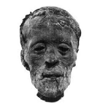
Focussing on the problem of facial reconstruction, we choosed a mummified
head in good condition, from the Egyptian Section of the Archaeological
Museum in Florence (inv. N. 8643). The date of its acquisition is 1893;
we lack any other reliable information about its provenance. C14 calibrated
dating of a sample of the hair gave a probability distribution between
339 b.C. and 201 b.C. [7]. The very good condition of
the head, attesting the quality of the embalming process, make us prefer
the higher dating.
The project involved five different stages:
1. anthropological and egyptological analysis of the head;
2. spiral CT of the head;
3. reconstruction of a 3-D model of the skull generated from CT data processing;
4. reconstruction of soft tissues;
5. application of textures fitting the somatic features.
The different stages are not strictly sequential: as we shall see, spiral
CT scannings and, later, their 3-D reconstruction provided new interesting
data to the previous phases (anthropological and egyptological investigations).
First phase of research: preliminary anthropological results
1. The anthropological study of the mummified cranial remains allowed us to identify a male subject with an age at death of around 40 years. The skull is dolichocranic, of medium height in norma lateralis, and with rounded occiput, narrow face, high cheekbones, gracile even if well developed in its height, jaw; the orbits are narrow, the nose is well-shaped, and of Europoid look.
The general appearance of the subject, especially regarding the face and the shape and structure of his hair, lead us to exclude Negroid influences, but closely resembles present and past Berber ethnic characters.
The very good conservation of the head pointed to an individual high in the social hierarchy, so as to grant himself an effective (and expensive) embalming process.
2. The mummy was scanned on 18th April 1997, using a Siemens Somatom Plus 4 spiral computer tomography scanner at Careggi Hospital in Florence, thanks to the kind collaboration of the radiological equipe. Slices thickness was 0.5 mm through all the skull.
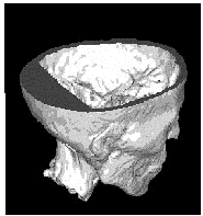 Fig. 2 CT scanning
of the head demonstrated the post-mortem transnasal ethmoid fracture created
by the embalmers to extract the brain tissue; the cranial cavity was filled
with hot melted resin, later solidified, introduced with the mummy resting
on its back, as the model reconstructed from the CT images clearly displays.
Fig. 2 CT scanning
of the head demonstrated the post-mortem transnasal ethmoid fracture created
by the embalmers to extract the brain tissue; the cranial cavity was filled
with hot melted resin, later solidified, introduced with the mummy resting
on its back, as the model reconstructed from the CT images clearly displays.Embalming excerebration through the ethmoid was very common in the Late
Period, practised until Ptolemaic age, as well as the filling of the cranial
cavity with resin [8].
The spiral CT images were later
electronically transferred to the Onyx2 workstation (Silicon Graphics)
at CINECA for post-processing.
3. In the methodology used for 3-D reconstructions generated by spiral CT data sets, CT slices must be stacked up and interpolated in order to build a volume. Once created a volume, it is possible, by means of suitable algorithms, to generate surfaces whose points have the same function value. They are called isosurfaces. A popular algorithm for determining isosurfaces is the so called marching cubes [9] , the same used in the 3-D reconstruction of our mummy?s skull. The principle underlying the application of this algorithm to the kind of problem here described is that similar materials have the same radio-opacity and are, consequently, represented in a CT scan by the same densitometric level. In CT slices, the intensity associated to each pixel in the grey-scale is proportional to tissues density: black corresponds to air, white to bones. It is therefore possible processing the CT scans sequence so as to obtain a 3-D grid, where to each "knot" (control point) is associated the densitometric value measured by the CT scans. The result is a 3-D 256 grey levels image.
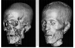
This phase of the work was particularly interesting from the anthropological
perspective: the use of this technique allowed us to visually exclude the
mummified soft tissues and directly observe the cranial bones with a very
high image resolution. This method offered also the chance of a morphological
and morphometric check of the anthropologist?s observations on the specimen
(covered by the disseccated soft tissue): these two methods of investigation
led to the same diagnosis of Berber group.
The image of the skull gave us the chance to observe, directly on the
cranial vault, a bone pathology and a biological answer to the pathogenic
factor that shows a long survival of the subject. X-ray examination of
the mummified skull showed in more detail the reactions to this kind of
pathology so that we could perform a global analysis of the sample.
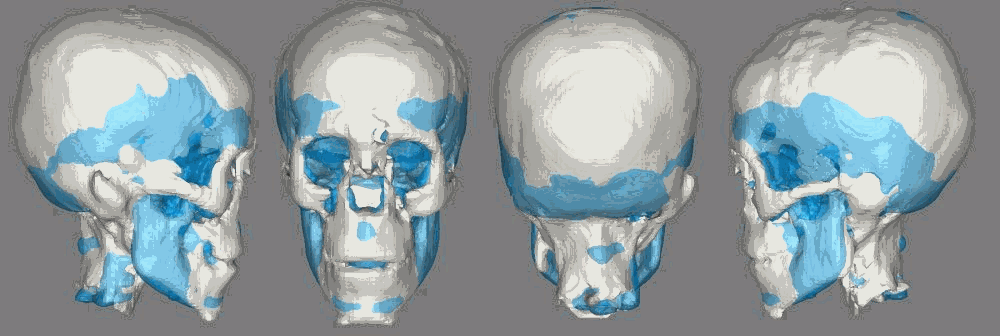
4. This stage of our work is still in a preliminary phase. Among the possible methodologies to deal with this complex problem, we focussed two different promising ways:
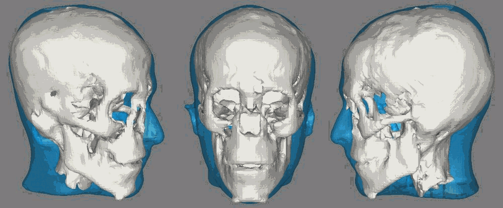
B. A different method consists in the distortion (warping) of
the 3-D model of a reference scanned head, until its hard tissues match
those of the mummy. The subsequent stage is the construction of the hybrid
model composed by the hard tissues of the mummy plus the soft ones of the
reference head [11].
We believe that very good results could outcome through a semiautomated
interactive procedure, integrating the two methodologies here described.
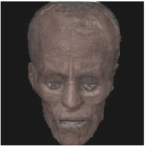
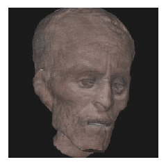
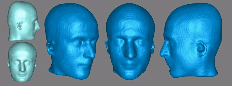
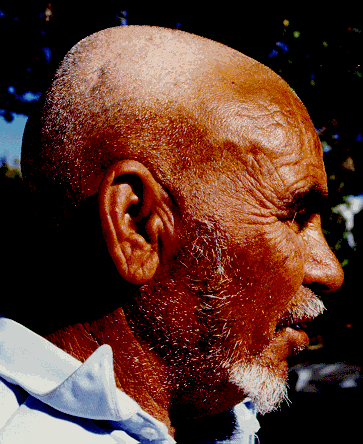
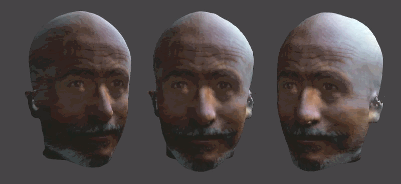
5. While hard and soft tissues give morphological information, textures provide colours and aesthetical features. They are "pasted" over the 3D models by means of mapping procedures. In this preliminary phase we used as texture the photograph of a modern Berber, published in an anthropological treatise [12], well fitting the general somatic features of our reconstruction but, unluckly, of very low resolution (fig. 4). Moreover, being a frontal view, it does not give sufficient information for the mapping of the entire model.
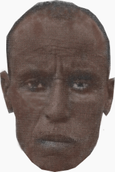
The texture was mapped onto the 3D model to perfectly match the frontal view of the mummy but it loses its grain as soon as we depart from the frontal view. Much better results could be obtained with different high resolution views of a new subject.
Development of the project: soft tissue reconstruction using VTK
After a first part of work, our open problem is to reconstruct the lacking
elements of a 3D digital model generated from CT scans applied to a mummified
cranial remains (Fig.7)
As we have described in the previous part of the paper, [14]
we work with an hybrid approach [13];
|
|
Soft tissues reconstruction
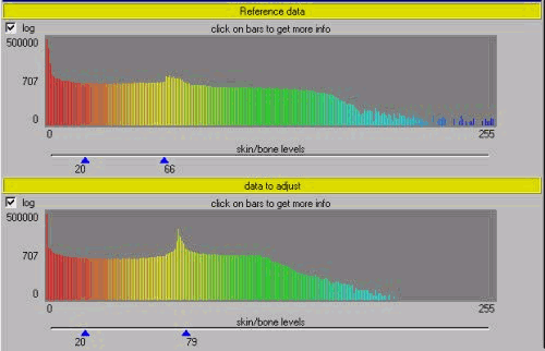
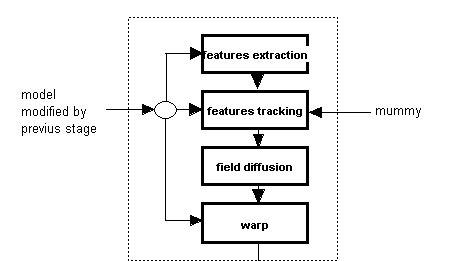
As shown in fig.8, our methodology may be subdivided in different working
steps that we are going to explain deeply.
|
|
 |
||
 |
 |
|
|
 |
 |
 |
 |
 |
 |
If ![]() is the intensity
of a point of coordinates (x,y,z) at time t in the mummy
volume and
is the intensity
of a point of coordinates (x,y,z) at time t in the mummy
volume and ![]() is the motion
field, where
is the motion
field, where ![]() ,
, ![]() e
e ![]() are components in x,
y e z directions of velocity vector, we suppose that the
intensity function is the same at the time
are components in x,
y e z directions of velocity vector, we suppose that the
intensity function is the same at the time ![]() in the point
in the point ![]() of the reference
model, where
of the reference
model, where ![]() ,
, ![]() e
e ![]() and
and ![]() .
.
(1)![]()
If the intensity function change smoothly with x, y, z e t, we can manipulate the equation (1) with Taylor?s series to obtain
(2) 
where e contains terms in dx, dy, dz e dt higher than first order.
Eliminating ![]() , rationing
by dt, and calculating limit for
, rationing
by dt, and calculating limit for ![]() ,
we obtain
,
we obtain
(3) 
that is the totally derivative of ![]() in the time.
in the time.
(4) ![]()
Using abbreviated notation:

we can write the 3 as
(5) ![]()
known as motion field constraint equation, where Ex, Ey, Ez ed Et are partial derivatives.
We say that x is a reliable feature if
(6) 
where:
I(![]() ,t) is the matrix
of intensity function E in the point
,t) is the matrix
of intensity function E in the point ![]() =(x,y,z)
in the region W(x) at the time t;
=(x,y,z)
in the region W(x) at the time t;
Ñ is the gradient operator;
s min(Y ) represents the smaller eigenvalue of matrix Y ;
![]() are predetermined thresholds.
are predetermined thresholds.
We consider a window ![]() (q)
centered in q of
(q)
centered in q of ![]() dimensions.
dimensions.
We represent (6) in discrete fashion
(7) 
where

The solution of (4) respect to V is given by
(8) 
where
![]()
is the 3x3 symmetric matrix that represents the term inside parenthesis of (7),
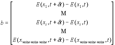
that is the intensity difference between reference and mummy volumes.
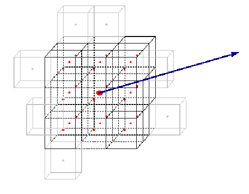
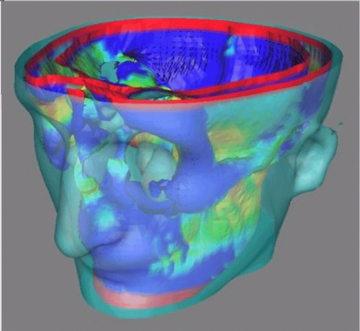
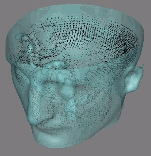
Normally this result is affected by an error of 10% approximately. To improve its precision we use an iterative multistep method: the window is moved in the mummy volume in the direction of estimated motion and the field is recalculated among new positions as shown in fig. 21.
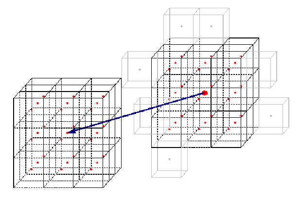
The process will be over when displacements become smaller than a predetermined
arbitrary constant or when a maximum limit to iterations is reached. In
case of non convergence the feature is discarded.
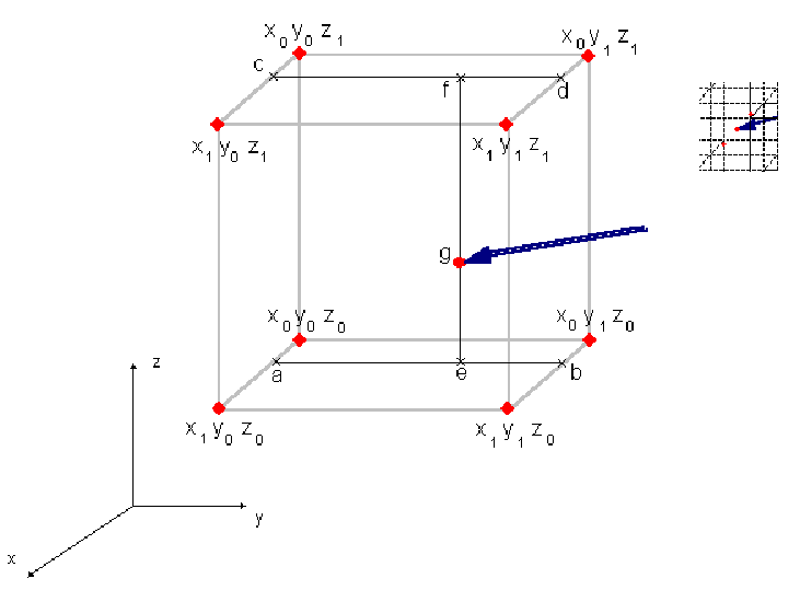
When a feature doesn?t coincide with a voxel, window points are calculated
by trilinear interpolation using vtk facilities (see fig. 22).
Texture application
We are now at summarizing mainly points of this stage, still under development,
inspired by the work presented in [22].
Starting from at least three views of our candidate, as in figure 23,
we will going to generate a cylindrical texture.
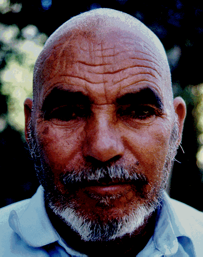
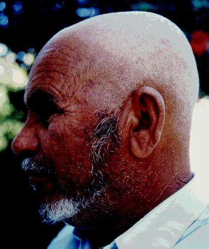
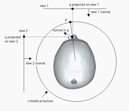
As shown in figure 24, each point P on the cylindrical texture will
be projected on the cylindrical axes, intersecting a point q on the surface,
watching the angle between the normal in q and the normal of the view we
can determine which of the views q can see,
The color for the Point P is given by a weighted sum of the colors
taken from the visible views, and the weights depend again on the angle
among the normals.
Before the extraction from the views colors has to be modified to match
the corresponding projection of the whole model (fig.25), we can realize
this morph implicitly after fixing a way to map projections in the
desired points of the view.
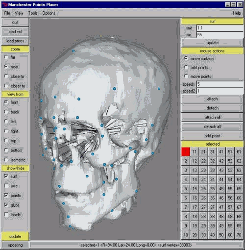
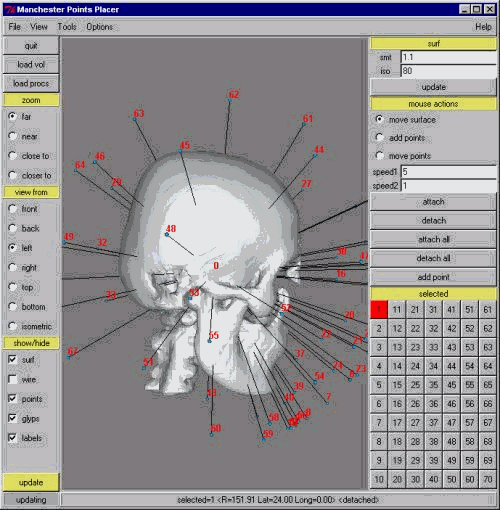
This mapping could be achieved by projecting the Manchester points
over the views and let an user move them in the right place.
 |
Conclusions: the virtual model
In our project, for obtaining better performances through the virtual
3D visualisation of the reconstruction we have used the powerful workstation
Onyx2 (Silicon Graphics at VISIT, CINECA), equipped with an architecture
of type multiprocessor, with 4 processors R10K, 1 Gbyte of RAM, computing
power of 1.5 Gflop, 1 graphic pipeline, that it can process 11 milions
of polygons per second. In fact the main problem, processing a large amount
of data, was to process and visualise in real time and in 3D the data volume.
The
interactive way to explore and show the whole model gives the possibility
to understand deep features of the data: in our case, our interdisciplinary
team, constituted by archaeologists, anthropologists, and computer experts,
has could discuss in real time problems and characteristics of the data
comparing in detail hypotesis and interpretations about the egyptian head,
anthropometric data and computer/images information. We can describe the
processing as a cognitive model of the find. Finally, we have experimented
the VR CrystalEyes, wireless pairs of glasses, with lenses capable of alternately
shuttering in sequence with left-eye right-eye images
interlaced on a computer display; the result is a stereoscopic effect,
allowing the user to view the contents of the computer display in 3D.
Possible develpoments of the project will concern:
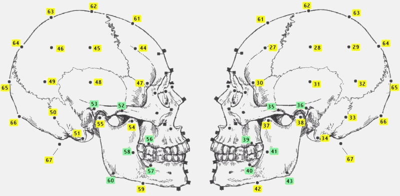
Appendix A: our set of skull markers
We have referred to the set of points illustrated onto tables by Rhine
& Moore . These points, originally is in number of 32 and mainly concentrated
on the face, are in green in figure 26.
For our purpose we add further points, the yellow ones (figure 27 )
and now we have a set of 67.
|
|
 |
Appendix B: detail for diffusing scattered fields and warp
Our building block to perform diffusion is the Shepard?s method that
is implemented by the vtkShepardMethod filter of the Vtk library.
Shepard?s method is an example of a basis function method , more specifically
is an inverse distance weighted interpolation technique that can be written
as:
To make this filter operate on vector values we write a new Filter that
internally instantiate three vtkShepardMethod and let them operate on the
single components as show in next figure.
|
|
References
| [1] | Petrie W.M.F., Deshasheh, 1897. Fifteenth memoir of the Egypt Exploration Fund. London 1898 |
| [2] | Notman, D., Ancient Scannings: Computed Tomography of Egyptian Mummies, in: ?Science in Egyptology. Proceedings of the ?Science in Egyptology Symposia? (ed. R.A. David)?, Manchester 1986, 251-320. |
| [3] | G. Foster, J.E. Connoll, J.Z. Wang, E. Teeter, P.M. Mengoni,
Evaluation of an ancient Egyptian Mummy using
spiral CT and 3-D reconstructions:interactive display using the World Wibe Web, Radiology 205(P), 734-35, 1997; on the web: http://www.rad.rpslmc.edu/rsnamumie/rsnamumie.html. |
| [4] | David A.R. (ed.), Proceedings of the ?Science in Egyptology Symposia?, Part I: Selected papers, 1979. Part II: papers presented 1984, Manchester 1986; Marx M., D?Auria S.H., Three-dimensional CT reconstructions of an ancient human Egyptian mummy, Amer. J. Roent. 150, 1988, 147-149; Drenkhahn R., Germer R. (eds.), Mumie und Computer. Ein multidisziplinäres Forschungsprojekt in Hannover, Sonderausstellung des Kestner-Museums Hannover, 1991; Baldock C., Hughes S.W., Whittaker D.K. et al., 3-D reconstruction of an ancient Egyptian mummy using x-ray computer tomography, Journal of the Royal Society of Medicine 87 (12), 1994 Dec., 806-808 (on the web:http://www-ipg.umds.ac.uk/MEDPHYS/projects/jen); G. Foster, J.E. Connoll, J.Z. Wang, E. Teeter, P.M. Mengoni, Evaluation of an ancient Egyptian Mummy using spiral CT and 3-D reconstructions:interactive display using the World Wibe Web, Radiology 205(P), 734-35, 1997; on the web: http://www.rad.rpslmc.edu/rsnamumie/rsnamumie.html. |
| [5] | See for example the pioneering work of Andreas Pommert
and Ulrich Kliegis in: Drenkhahn R., Germer R. (eds.),
Mumie und Computer. Ein multidisziplinäres Forschungsprojekt in Hannover, Sonderausstellung des Kestner-Museums Hannover, 1991; or the impressive reconstruction of a young child?s face in the University of Illinois mummy project: Sarah Wisseman et al., Imaging the Past..., in: Ancient Technologies and Archaeological Materials, 1994, 217-234 (on the web: http://www.grad.uiuc.edu/departments/ATAM/imaging.html. Also in the facial reconstruction of a sailor whose remains were found during the archaeological exploration of the La Salle shipwreck, three-dimensional imaging of the skull was used to generate an exact model of the head through stereolithography; the following stage was the construction -by an artist - of a clay model of the face, based on anthropological tissue measurements. |
| [6] | F. Mallegni, Un volto di gioventù per il principe
Uage, in: M. Forte (ed.), Archeologia. Percorsi virtuali nelle civiltà
scomparse, Milano 1996, 50. |
| [7] | The analysis was effectuated in the ANSTO Laboratory, Menai, Australia. Ref. Code 0ZD005. |
| [8] | Strouhal E., Embalming excerebrtion in the Middle kingdom,
in: R. David (ed.), Science in Egyptology, Manchester
1986, 141-154; Id., Secular Changes of Embalming methods in Ancient Egypt, in: Actas del I Congreso Internacional des estudios sobre momias, Tenerife 1992, 862-63; Macke A., Les orifices d?viscérations endocraniennes aux Basses Epoques, Annales du Service des Antiquités Egyptiennes 72 (1993), 135-147. |
| [9] | W.E. Lorensen and H.E. Cline, Marching Cubes: A high
resolution 3D surface construction algorithm, in: Proc.
SIGGRAPH ?87 (Anaheim, Calif., July 27-31, 1987), vol. 21(4), pp. 163-169. ACM SIGGRAPH, New York July 1987. |
| [10] | Human Skeleton Remains, Washington 1989, table 122-123. |
| [11] | T. Beier and S. Neely, Feature-based image metamorphosis, Computer Graphics, vol 26(2), pp 35--42, New York, NY, July 1992. Proceedings of SIGGRAPH '92; P. Bergeron. Morphing as a means of generating variation in visual medical teaching materials. Computers in Biology and Medicine, 24(1):11--18, Jan. 1994; A. Kaufman, D. Cohen, and R. Yagel. Volume graphics, Computer, 26(7):51--64, July 1993; P. Litwinowicz. Efficient techniques for interactive texture placement, Computer Graphics Proceedings, Annual Conference Series, pp 119--122, New York, NY, July 1994. Conference Proceedings of SIGGRAPH '94; P. A. van den Elsen, E.-J. D. Pol, and M. A. Viergever, Medical image matching --- a review with classification, IEEE Engineering in Medicine and Biology Magazine, 12(1):26--39, Mar. 1993; G. Wolberg, Digital Image Warping. IEEE Computer Society P., Los Alamitos, CA, 1990; S.D. Michael and M. Chen, The 3D Reconstruction of Facial Features Using Volume Distortion, Proceedings of Eurographics (UK Chapter) (eds. H. Jones, R. Raby and D. Vicars), ISBN 0 952 1097 5 1 (Vol. 2) (1996), pp. 297-305. |
| [12] | R. Biasutti, Razze e popoli della Terra?, vol. 3, 130. |
| [13] | M. Betrò, M. Forte, R. Gori, F. Mallegni and A. Sarti. "3D facial reconstruction and visualization of ancient Egyptians mummies using spiral CT data" |
| [14] | Will Schroeder, Ken Martin, Bill
Lorensen "The Visualization Toolkit" 2nd.Edition Prentice Hall
PTR
http://www.kitware.com |
| [15] | Frederic Pighin, Jamie Hecker, DaniLischinski, Richard Szeliski, David H.Salesin "Synthesizing Realistic Facial Expressions from Photographs" SIGGRAPH 98 Proceedings |
| [16] | B. Lucas and T. Kanade. "Performance of otpical flow techniques" proc. DARPA IU Workshop 1981. |
| [17] | C. Lamberti, A. Sarti and F. Fraticelli, "Computation of 3D velocity fields from 3D of echocardiographies of human heart." |
| [18] | AIR: Automatic ImageRegistration http://bishopw.loni.ucla.edu/AIR/ |
| [19] | John Prag and Richard Neave Making
Faces Using Forensic and Archaeological Evidence
http://www.britarch.ac.uk/ba/ba31/ba31book.html |
| [20] | Table of measurements for flesh thickness, after J.S.Rhine and C.E.Moore" Forensic Anthropology, Maxwell Museum Technical Series 1 (1984) |
| [21] | J.Wixom and W.J.Gordon "On Shepard?s Method of Metric Interpolation to Scattered Bivariate and Multivariate Data" Math. Comp. 32:253-264, 1978 |
| [22] | Morten Bro-Nielsen "Medical image registration and surgery simulation" Ph.D. thesis at technical University of Denmark (1996) |
Special thanks are due to the CINECA-Visit (Visual Information lab), to the Careggi Hospital (Florence) and to the Archaeological Museum of Florence for the fundamental support in our research project.